Authors:
Belaid L1, Rivalan J1, Laguerre B2, Rioux-Leclercq N3,4 and Vigneau C1-3*
1CHU Pontchaillou, service de néphrologie, Rennes, France
2CRLCC, Rennes, France
3Université Rennes 1, CNRS UMR 6290, équipe Kyca, faculté de Médecine, Rennes, France
4CHU Pontchaillou, service d'anatomie et cytologie pathologiques, Rennes, France
Received: November 04, 2014; Accepted: January 08, 2015; Published: January 10, 2015
C Vigneau, CHU Pontchaillou, service de néphrologie, 2 rue H Le Guilloux 35000, Rennes cecile, Email:
Belaid L, Rivalan J, Laguerre B, Rioux-Leclercq N, Vigneau C (2015) An Anusual Cause of Secondary Hypertension in a Young Man. J Cardiovasc Med Cardiol 2(1): 017-018. 10.17352/2455-2976.000011
© 2015 Belaid L, et al. This is an open-access article distributed under the terms of the Creative Commons Attribution License, which permits unrestricted use, distribution, and reproduction in any medium, provided the original author and source are credited.
Case Report
Mr F.P., a 33-y/o man consulted after 3 months of general status alteration with a loss of 7 kgs and nocturnal sweats. His blood pressure was controlled up to 190/110 mmHg. Clinical examination by general pratictioner was considered as normal. Bitherapy by calcium channel blockers and alpha blockers was given.
Few days later, hypertension was still severe between 160/100 and 200/120mmHg despite the bitherapy. Blood analysis revealed potassium at 3,1mmol/L. The patient was then hospitalized in a nephrological department. Plasma renin activity and aldosterone were elevated as shown on (Table 1). Clinical examination revealed a 8 cm mass in the left testicul, painless, of tissular consistency, without any inflammatory sign or adenopathy. Mr FP has noticed this mass for one year but this was rapidly growing within the last 2 months. Ultrasound and CT scan showed that the mass was depended on the left spermatic cord and associated with one nodule on liver, two juxta-hepatic, an osteolysis on the right hip and a mediastinal lymph node mass. The biopsy of the liver nodule revealed a leiomyosarcoma with high malignity staging.
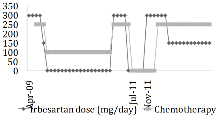
Figure 1 shows the clinical evolution under therapeutics. An angiotensin receptor blocker (irbesartan 300mg per day) was introduced in april 2009 in place of bitherapy and succeeded to normalize both blood pressure and kaliemia (respectively 130/80mmHg and 4,5mmol/L). A chemotherapy by taxotere-gemcitabine was introduced in may 2009. After 6 cycles, the tumor was reduced on CT-scan. Blood pressure was low (100/60mmHg) leading to irbesartan interuption with normal bood pressure (120/70mmHg) afterwards. Maintenance chemotherapy was sustained with gemcitabine.
-

Figure 1
Evolution of daily dose of irbesartan and chemotherapies (docetaxel gemcitabine high dose for intensive treatment and gemcitabine low dose for maintenance therapy).
In february 2011, blood pressure increased again and patient reported left hip pain. CT scan showed an aggravation of the osteolysis. Gemcitabine and irbesartan were restarted. After 6 additional cycles of taxotere-gemcitabine, CT scann showed partial response, blood pressure was low and therapeutics (taxotere-gemcitabine and irbesartan) could be again interupted.
In november 2011, the patient was hospitalized in emergency for high blood pressure and hip pain. He benefited of analgesic radiotherapy and treatment by irbesartan 300mg per day.
In february 2012, chemotherapy with 4 cycles of taxotere-gemcitabine was restarted and the daily dose of irbesartan can be decreased to 150mg. However, response was only partial and another chemotherapy by 3 cycles of doxorubicine-dacarbazine failed to improve osteolysis. Finally, another chemotherapy by yondelis induced stability of bone lesions on CT-scann and irbesartan dose can be again reduced.
Discussion
This case is the first described of paraneoplasic hypertension revealing testicular tumor. This case is of particular interest since evolution over 3 years, shows a perfect correlation between blood pressure and leiomyosarcoma evolutivity. Moreover, since the patient understood this link, blood pressure automeasure were able to help detecting early tumor relapses.
Mr FP presented with secondary hypertension, with hyperreninism which can be due to paraneoplasic syndrom or renin secretion by the tumor. Because of metastasis, orchidectomy was not indicated and no primary tumor histology could be realized. Liver metastasis biopsy did not show any staining for renin but we cannot eliminate a primary renin tumor in testicule.
Renin secreting tumors are rare. The most frequent histological type is renal juxta-glomerular apparatus tumor [4]. However, uterin leiomyosarcoma has already been described in a 60 y/o woman with hypertension and hypokaliemia [1]. Immunocytochimy revealed renin expression. Blood pressure got normal after tumor removal. Hyperreninism has also been described with ovarian Sertoli cell tumor [2] and neuroendocrine pancreatic carcinoma [3]. Finally, this case underlines the importance of a complete examination and a biological assessment in front of unusual hypertension, especially if hypertension is resistant and/or on a young patient. Role of hormones on vascular tone and blood pressure should never be negliged as well showed on postmenopausal women treated with estrogeno therapy [5,6].
- Moody AM, Ahmed A, Chakrabarty A, Fisher C (1995) Renin-producing leiomyosarcoma originating in the uterus. Clin Oncol 7: 52-53
- Korzets A, Nouriel H, Steiner Z, Griffel B, Kraus L, et al. (1986) Resistant hypertension associated with a renin-producing ovarian Sertoli cell tumor. Am J Clin Pathol 85: 242-247.
- Langer P, Bartsch D, Gerdes B, Schwetlick I, Wild A, et al. (2002) Renin-producing neuroendocrine pancreatic carcinoma-a case report and review of the literature. Exp Clin Endocrinol Diabetes 110: 43-49.
- Wong L, Hsu THS, Pelroth MG, Hofmann LV, Haynes CM, et al. (2008) Reninoma: Case report and literature review. J Hypertension 26: 368-373.
- Ciccone MM, Scicchitano P, Cortese F, Gesualdo M, Zito A, et al. (2013) Modulation of vascular tone control under isometric muscular stress: role of estrogen receptors. Vascul Pharmacol 58: 127-133.
- Ciccone MM, Cicinelli E, Giovanni A, Scicchitano P, Gesualdo M, et al. (2012) Ophthalmic artery vasodilation after intranasal estradiol use in postmenopausal women. J Atheroscler Thromb 19: 1061-1065.


Table 1:
Biochemical parameters.
Patient
Norm
3,1
3,5-5,0
413
1925
10-100
30-270
> 320
> 320
10-48
14-71
Table 1: Biochemical parameters.