Authors:
Saime Güzelsoy Sagiroglu1*, Hüseyin Öztarakci2 and Adem Doganer3
1Department of Otorhinolaryngology, Faculty of Medicine, Kahramanmaras Sutcu Imam University, Kahramanmaras, Turkey 2Department of Otorhinolaryngology, Kahramanmaras Necip Fazil Sehir State Hospital, Kahramanmaras, Turkey
2Department of Biostatistics and Medical Informatics, Kahramanmaraş Sütçü İmam University, Faculty of Medicine, Kahramanmaraş, Turkey
Received: 12 January, 2017; Accepted: 25 January, 2017; Published: 28 January, 2017;
Saime Güzelsoy Sagiroglu, Department of Otorhinolaringology, Faculty of Medicine, Kahramanmaras, 46100, Turkey, Tel: +90 344 2803335; +90 505 2400544; Fax: +90 344 221 2371; E-mail:
Sagiroglu SG, Öztarakci H, Doganer A (2017) The Evaluation of Hematologic and Platelet Function in Total Septum Reconstruction Patients. Arch Otolaryngol Rhinol 3(1): 017-021. DOI: 10.17352/2455-1759.000036
© 2017 Sagiroglu SG, et al. This is an open-access article distributed under the terms of the Creative Commons Attribution License, which permits unrestricted use, distribution, and reproduction in any medium, provided the original author and source are credited.
Objective: Nasal septal deviation is a common disorder of the nose and patients commonly visit clinics with complaints of nasal obstruction. As a result of nasal obstruction, patients are exposed to snoring, which causes hypoxia. Hypoxia leads to chronic inflammation, oxidative stress and endothelial dysfunction. Platelet-to-lymphocyte ratio (PLR) and neutrophil-to-lymphocyte ratio (NLR) are new markers for inflammation. In this study we have aimed to study the relationship between hypoxia, PLR, NLR and other platelet markers in patients with septum and external deviations.
Methods: 100 patients (77 male, 23 female) with nasal obstruction, external nasal deformity and snoring symptoms, with severe septum deviations and external deviations were studied. The control group consisted of 101 (73 male, 28 female) people, making it in total 201 people in the study. Preoperative blood samples were taken from these subjects to look at the Hb (haemoglobin), Htc (haematocrit), RBC (the number of red blood cells), MCV (mean corpuscular volume), MCH (mean corpuscular haemoglobin), MCHC (mean corpuscular haemoglobin volume), MPV (mean platelet volume), neutrophils, lymphocytes PDW (platelet distribution ratio), PC (platelet count), PLR and NLR values.
Results:The PLR and NLR values were significantly higher in the control group compared with the female patient group. The PDW and MCH values were significantly higher in the male patient group compared with the control group.
Conclusion:We have shown that PLR and NLR values is a correlation in female patients with septum deviation. And we detected that PDW values were high in male patients with septum deviation. Patients with apparent septum deviation should be subject to surgery as soon as possible.
Introduction
Nasal obstruction is one of the most common complaints within the otolaryngology clinics and it is mainly caused by septum deviation [1,2]. Septum deviation has a close relationship with hypoxia and with hypoxia average platelet counts increase [3,4]. Platelets have an important role in damage repair in both endothelial and haemostasis. Platelets produce proinflammatory mediators such as cytokines and chemokine’s during vascular inflammation. Furthermore, they also lead to release of mediators from the cells in the vascular wall. Activate platelets lead to formation of thrombi by either repairing the endothelial cell erosion or forming an atherosclerotic plaque. This triggers the start of atherothrombotic diseases [5,6].
Snoring and hypoxia caused by nasal obstruction is usually caused by septal and external deviation, which is treated surgically. Rhinoplasty is technique applied in two ways as closed and open (external), and it allows visibility into the nose cartilage and bone structure, also allows bimanual surgery manipulations and grafting [7]. The first explanation of rhinoplasty is from Indian Sushutra and Samhita from MO 600. Open rhinoplasty was introduced by Rheti in 1934 [8,9].
With this study we have aimed to demonstrate the hemogram values, platelet marks, PLO and NLO values which have been changed due to hypoxia in patients who have gone under septal reconstruction by open rhinoplasty.
Materials and Methods
This is a retrospective study consisting of patients with nasal congestion, snoring and misshaped nose symptoms, with severe septal deviations and external deviations who have visited the clinic between 2013-2016. The patient group consisted of 77 males and 23 females (total 100), with mean age of 22.5 ±4.7 (18-34). The control group was consisted of healthy volunteers with similar ages, 73 males and 28 females (total 101) with a mean age of 22.11±3.5 (18-32). Inclusion criteria control group was not presenting with complaints of nasal obstruction, snoring which on anterior rhinoscopy and nasal endoscopy were not diagnosed to have deviated nasal septum.
The patients with marked septal deviation were diagnosed by anterior rhinoscopy and nasal endoscopy (Karl Storz, Germany). A lot of patients had a nasal trauma story. The other problems causing upper airway obstruction such as hypertrophied tonsils, adenoid vegetation, nasal polyposis and chronic rhinosinusitis were excluded in this study. The patients with rhinosinusitis and nasal polyps were followed up with medical treatment and not taken in to the study. There weren’t apnea stories in patients. The patients consisted of simple snorers but all patients were extremely hard breathing. Each of the cases has had open rhinoplasty. All the patients had at least one nasal passage completely blocked due to caudal deviation and severe septal deformities. As a result, the septum was completely removed and extracorporeal septum reconstruction was applied. Lateral osteotomy was applied to some patients following a hump excision. The preoperative blood samples were transferred to tubes containing ethylene-diamine-tetraacetic acid (EDTA). Venous blood samples were used to measure Hb (haemoglobin), HTC (haematocrit), RBC (red blood cell count), MCV (mean corpuscular volume), MCH (mean corpuscular haemoglobin), MCHC (mean corpuscular haemoglobin volume), MPV (mean platelet volume), neutrophils, lymphocytes PDW (platelet distribution ratio) and PC (platelet count). Blood counts were studied by an auto-analyser (Abbott Cell-Dyn 3700 Hematology Analyzer, USA).
Statistics
In the patient group n=100 and control n=101 cases were evaluated. The male and female were done separately the statistical analysis. The statistical analysis was completed using SPSS 20 programme. Unpaired two-sample t-test is used to analyse results. For results showing normal distribution parametric tests were used. Non-parametric tests were used for the analysis of results that did not fit normal distribution. For results that fit normal distribution, the control group and the patient groups values were compared using unpaired two-sample t-test, for the age comparisons one-way analysis of variance (ANOVA) was used. For the descriptive analysis mean and standard deviation was used. For the values that did not show normal distribution, a Mann-Whitney U test was used to compare the patient and control groups. To compare the ages, Kruskal Wallis H test was used. For the descriptive statistics the median (minimum-maximum) was used. p<0.05 was used for statistically significant.
Results
Present study includes 100 cases, out of which, 77cases (51.3 %) were males while 23 cases (45.1 %) were females. Among 101controls, 73 (48.7 %) were males and 28(54.9 %) females. The patient group consisted of 77 males and 23 females, with mean age of 22.90±4.72 and 21.17±4.56. The control group consisted of 73 males and 28 females, with mean age of 22.67±3.70 and 20.68±2.51. Average Hb, Htc, RDW, lymphocyte, neutrophil, PC, RBC, MCH, PDW, MCHC, MPV, PLR, NLR values are presented on Tables 1,2. The male patient group showed significantly higher PDW and MCH values compared to the control group (Chart 1,2). The incidence of the male patient in PLR and NLR was also studied but the observation was statistically not significant. According to the analysis, PLR and NLR parameters between the female patients and control groups were found to be statistically significant (p=0.039, p=0.017) (Chart 3,4). Furthermore, the lymphocyte %, neutrophil, neutrophil % parameters differences were also found to be statistically significant between the female patient and control group (p=0.015, P=0.027, p=0.025) (Chart 5,6). The patients that no significant differences were found between PC, RBC, MCHC, MPV, Hb, Hct and RDW values. The smoking group included 58 (%28.9) and non-smoking group included 143 (% 71.1). The recorded level of smoking among male patients was 42 (%28). The recorded level of smoking among female patients was16 (%31.4). There was no significant difference in the presence smoking between the two groups (p=0.328).
-
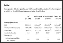
Table 2:
Comparison of parameters for patient and control group (Female).
-
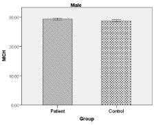
Chart 1:
MCH value(male).
-
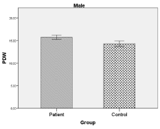
Chart 2:
PDW value (male).
-
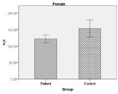
Chart 3:
PLR value (female).
-
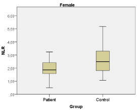
Chart 4:
NLR value (female).
-
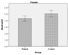
Chart 5:
Neutrophil value (female).
-
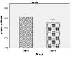
Chart 6:
Lymphocyte radio(%)(female).
Discussion
Nasal obstruction is one of the most common symptoms in nasal septum deviation [1,2,10]. Septum deviation accompanied by the nasal roof, is less commonly seen than insulating septum deviations. Due to nasal obstruction, patients are exposed to intermittent hypoxia. One of the consequences of hypoxia is the difference in platelet function. As shown by previous studies, chronic hypoxia could lead to many cardiovascular diseases by the activation of platelets leading to hypercoagulation (7,11,12). Similar platelet activation mechanisms are seen in nasal obstruction, which is one of the causes of intermittent hypoxia.
MPV is a parameter that is used in evaluating the size of platelets, as well as being a marker for platelet reactivation potential [3,13]. MPV is a routine hemogram parameter and it gives information on platelet function. Larger platelets are more adhesive and are inclined to be more aggressive [5,6,14]. MPV increases with age, while platelet count decreases [14]. Some studies have shown serious increases in MPV values in obstructive sleep apnea syndrome (OSAS) patients who have developed chronic hypoxia due to snoring [5,13,15]. Cengiz et al. [13], have discovered a negative correlation in MPV values in patients with chronic hypoxia who are suffering from chronic tonsilitis-adenoid hypertrophy. In our study, MPV values were found to be in normal range and the differences were not statistically significant.
PLR is a new biomarker, which allows detecting inflammation in both patients with cardiac or noncardiac diseases [11,16]. Song et al. [11], have claimed that PLR and PDW values are inflammatory markers that can show platelet activation and inflammation. Several studies have also shown that PLR and NLR values are markers that detect inflammation [7,12,17,18]. In our study we have found that the PLR and NLR values were not significant in male patients. We have found that PLR and NLR values were significantly in female patients group. Koseoglu et al. [5], have studied MPV and PLR values in OSAS patients and have shown that as the severity of OSAS increased, the PLR values also increased. They also suggested that PLR could be used as a biomarker in OSAS patients with cardiovascular diseases. In patients suffering from septum deviation, it was seen that as age increased, MPV and PDW values increased, which also increased the risk of cardiopulmonary diseases [3]. The male patient group showed significantly higher PDW. Therefore, we can say that the first changes in platelets due to hypoxia start with PDW and that PDW may be a good criterion.
This study showed that, within the young male patient population, septum deviation accompanied by the nasal roof results, the first change in PDW value being an increase. We detected that PLR and NLR values is a significantly in young female patient. As nasal obstruction period is elongated, also increase the risk of cardiopulmonary complication risk in patients. Concerning the mechanisms that are responsible for the age-related changes, this may be effect a variation in hematopoietic stem cell reserve during aging. However, the time that cardiopulmonary complication will occur is not known; patients with apparent septum deviation should be subject to surgery as soon as possible. To conclude, studies have shown that markers, which show the evaluation of hematologic, are also markers that lead in the mortality and morbidity of diseases, which develop due to snoring in patients with septum and external deviations. We believe that further retrospective and prospective studies are required for the markers, which show hematologic functions and platelet activation.
Ethics Committee Approval
The study was approved by the Local Ethic Committee of Kahramanmaras Sutcu Imam University, 70-03/2016.
-
-
- Haack J, Papel ID (2009) Caudal Septal Deviation. Otolaryngol Clin N Am 42: 427–436. Link: https://goo.gl/EBPyhf
- Moore M, Eccles R (2011) Objective evidence forthe efficacy of surgical management of the deviated septum as a treatment for chronic nasal obstruction: a systematic review. Clin Otolaryngol 36: 106-113. Link: https://goo.gl/DqjNrI
- Unlu I, Kesici GG, Oneç B, Yaman H, Guclu E (2016) The effect of duration of nasal obstruction on mean platelet volume in patients with marked nasalseptal deviation. Eur Arch Otorhinolaryngol 273: 401-405. Link: https://goo.gl/fqjan4
- Sagit M, Korkmaz F, Kavugudurmaz M, Somdas MA (2012) Impact of septoplasty on mean platelet volüm elevels in patients with marked nasal septal deviation. J Craniofac Surg 23: 974-976. Link: https://goo.gl/JGMVFW
- Koseoglu HI, Altunkas F, Kanbay A, Doruk S, Etikan I, et al. (2015) Platelet-lymphocyteratio is an independent predictor for cardiovascular disease in obstructive sleep apnea syndrome. J Thromb Thrombolysis 39: 179-185. Link: https://goo.gl/T9bjMy
- Wedzicha JA, Cotter FE, Empey DW (1988) Platelet size in patients with chronic airflow obstruction with and without hypoxaemia. Thorax 43: 61-64. Link: https://goo.gl/bqyUu4
- Varım C, Varım P, Acar BA, Vatan MB, Uyanık MS, et al. (2016) Usefulness of theplatelet-to-lymphocyteratio in predicting the severity of carotid artery stenosis in patients undergoing carotid angiography. Kaohsiung J Med Sci 32: 86-90. Link: https://goo.gl/B2ujDP
- Vuyk HD, Kalter PO (1993) Open septorhinoplasty. Experiences in 200 patients. Rhinology 31: 175-182. Link: https://goo.gl/R6M0a8
- Chaaban M, Shah AR (2009) Open Septoplasty: Indications and Treatment. Otolaryngol Clin N Am 42: 513–519. Link: https://goo.gl/ZoZfp2
- Moghadas H, Abouali O, Faramarzi A, Ahmadi G (2011) Numerical investigation of septal deviation effect on deposition of nano/microparticles in human nasal passage. Respir Physiol Neurobiol 177: 9–18. Link: https://goo.gl/cIWACk
- Song YJ, Kwon JH, Kim JY, Kim BY, Cho KI (2016) Theplatelet-to-lymphocyte ratio reflects the severity of obstructive sleep apnea syndrome and concurrent hypertension. Clin Hypertens 22: 3-8. Link: https://goo.gl/omZwBC
- Chen SC, Lee MY, Huang JC, Tsai YC, Mai HC, et al. (2016) Platelet to Lymphocyte Percentage Ratio Is Associated With Brachial–Ankle Pulse Wave Velocity in Hemodialysis. 95: 1-5. Link: https://goo.gl/wPltJj
- Cengiz C, Erhan Y, Murat T, Ercan A, Ibrahim S, et al. (2013) Values of mean platelet volume in patients with chronic tonsillitis and adenoid hypertrophy. Pak J MedSci 29: 569-572. Link: https://goo.gl/1CSf7y
- Poorey VK, Thaku P (2014) Effect of Deviated Nasal Septum on Mean Platelet Volume: A Prospective Study. Indian J Otolaryngol Head Neck Surg 66: 437–440. Link: https://goo.gl/IVJIYb
- Varol E, Ozturk O, Gonca T, Has M, Ozaydin M, et al. (2010) Mean platelet volüme is increased in patientswith severe obstructive sleep apnea. Scand J ClinLabInvest 70: 497-502. Link: https://goo.gl/YhfJU4
- Dotsenko O, Chaturvedi N, Thom SA, Wright AR, Mayet J, et al. (2007) Platelet and leukocyte activation, atherosclerosis and inflammation in Europeanand South Asian men. J Thromb Haemost 5: 2036–2042. Link: https://goo.gl/OSeMVz
- Geiser T, Buck F, Meyer BJ, Bassetti C, Haeberli A, et al. (2002) Invivo platelet activation is increased during sleep in patients with obstructive sleep apnea syndrome. Respiration 69: 229-234. Link: https://goo.gl/J7ZCHS
- Meng X, Wang W, Wei G, Chang Q, He F, et al. (2016) A highor a reasonably-reactively elevated plateletto-lymphocyteratio, which plays the role? Platelets 27: 491-495. Link: https://goo.gl/XqIKdE
-
-
-


Table 1:
Comparison of parameters for patient and control group (Male).