Authors:
William Lawson1, Anthony Reino2 and Robert Deeb3*
1Department of Otolaryngology, Head and Neck Surgery, Mount Sinai School of Medicine (WL), New York, USA
2Department of Otolaryngology, Head and Neck Surgery, James J Peterson Veterans Affairs Medical Center (AR), Bronx, New York, USA
3Department of Otolaryngology, Head and Neck Surgery, Henry Ford Health System, Detroit, Michigan (RD), USA
Received: 13 July, 2017; Accepted: 17 August, 2017; Published: 18 August, 2017
Robert Deeb, MD, Department of Otolaryngology-Head and Neck Surgery, Henry Ford Hospital, 2799 West Grand Boulevard, Detroit, MI, 48202, USA, Tel: 313-916-3272; Fax: 313-916-7263; E-mail:
Lawson W, Reino A, Deeb R (2017) The Riedel Procedure - An Analysis of 22 Cases. Arch Otolaryngol Rhinol 3(3): 087-092. DOI: 10.17352/2455-1759.000054
© 2017 Lawson W, et al. This is an open-access article distributed under the terms of the Creative Commons Attribution License, which permits unrestricted use, distribution, and reproduction in any medium, provided the original author and source are credited.
Riedel procedure; Frontal sinus osteomyelitis
Objectives: To report one institution’s experience with 22 cases of the Riedel procedure in order to establish a profile for those patients with chronic frontal sinusitis who develop chronic osteomyelitis.
Study Design: Retrospective review.
Methods: Review of all patients undergoing the Riedel procedure at one institution with analysis of demographic data, indications for procedure, technical aspects of performing the procedure, outcomes and complications. Comprehensive review of the literature was performed as well.
Results: Twenty-two patients were identified. The age range in patients varied from 20 years to 86 years. The etiology of the condition requiring the Riedel procedure was infectious in 16 patients, traumatic in 5 and neoplastic in 1. All patients with chronic osteomyelitis of the frontal sinus had undergone multiple intranasal and external procedures. Post-operative follow-up interval ranged from 4 months to 21years.
Conclusion: The Riedel procedure remains a relevant tool in the armamentarium of the rhinologic surgeon. Its primary indication, chronic frontal osteomyelitis, is an indolent process that generally occurs as sequelae of Pott’s Puffy tumor, trauma or neoplasm. The natural history is that of multiple failed endoscopic and open procedures. Despite the radical nature of the Riedel procedure, it may not be curative and patients require long term follow up.
Introduction
The Riedel procedure is a radical ablative operation devised in 1898 when, in the pre-antibiotic era, acute and chronic frontal sinusitis carried a high morbidity and mortality. Curiously it persists as part of the armamentarium of the sinus surgeon over a century later. Despite all the advances made in modern therapeutics, imaging and instrument technology, a small subset of patients persists with chronic refractory frontal osteomyelitis that has defied conventional management. It is the purpose of this paper to report our experience with 22 cases undergoing the Riedel procedure to establish a profile for those patients with chronic frontal sinusitis who insidiously develop chronic osteomyelitis and ultimately require radical ablative surgery.
Materials and Methods
A comprehensive chart review of all patients presenting to the senior authors (W.L. and A.R.) requiring the Riedel procedure was performed. Basic demographic information was recorded. The entire pre-operative, operative and post-operative course of all patients was reviewed including previous procedures and complications. A comprehensive literature review of frontal osteomyelitis and its management was performed as well.
Results
A retrospective review of the records at the Mount Sinai Medical Center, New York, NY, revealed 22 patients who had undergone the Riedel procedure in the period of 1975 to 2001 by the senior authors (W.L and A.R.). The follow-up interval ranged from 4 months to 21 years with a mean of 6 years. Table 1 represents a summary of the cases. Twenty males and 2 females with an age range of 20 to 86 years comprised the study group. Among 20 patients with chronic osteomyelitis, the mean age was 51, with a bimodal distribution (20-40 years in 10 cases, 55-86 years in 10 cases). Fifteen patients presented with refractory osteomyelitis and Pott’s puffy tumor and fistulae secondary to chronic infection of the frontal sinus (Figure 1). Five patients had sustained severe trauma to the frontal sinus secondary to motor vehicle accidents and gunshot wounds and subsequently developed chronic osteomyelitis of the frontal bone (Figure 2). The interval between the injury and the onset of osteomyelitis was generally prolonged ranging from 2 to 25 years. One patient had squamous cell carcinoma of the frontal sinus. All patients with chronic osteomyelitis of the frontal sinus had undergone multiple intranasal and external procedures as listed in table 2.
-
Table 2:
Procedures Performed Before Frontal Collapse in 21 Patients.
-
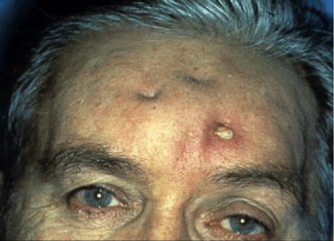
Figure 1:
Photograph of patient presenting with a sinocutaneous fistula secondary to chronic infection of the frontal sinus.
-
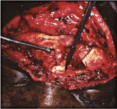
Figure 2:
Intraoperative view of patient who developed chronic osteomyelitis secondary to trauma.
In the 16 patients with chronic frontal sinusitis, 15 had at least 1 intranasal/endoscopic sphenoethmoidectomy and frontal sinusotomy. Four patients had an external frontoethmoidectomy, 1 had a trephine procedure, 10 had osteoplastic obliteration and 4 patients had a previous craniotomy (Table 1). One of each of the 5 traumatic cases had a repair of laceration, craniotomy, external frontoethmoidectomy, frontal exploration and osteoplastic flap with craniotomy (Table 2). A summary of the operative procedures performed in these cases is found in table 2.
Discussion
Among the paranasal sinuses, the frontal sinus presents the greatest challenge. It is the sinus most prone to multiple surgical procedures, both endoscopic and external, for a variety of reasons including: 1) maintaining nasofrontal outflow patency is limited by the tendency to osteoneogenesis; 2) mucociliary flow is from the nasal cavity into the frontal sinus inviting reinfection; 3) anatomically, diploe of the anterior and posterior table invites the development of osteomyelitis, and the diploic veins passing through them may lead to subperiosteal and intracranial infections; 4) it has the greatest incidence of mucoceles and mucopyoceles, which may indolently produce chronic infection of the surrounding bone; and 5) it has a longer and irregular outflow tract narrowed by extramural ethmoid cells.
Consequently, despite the increased safety and outcome results of endoscopic frontal sinus surgery enhanced by navigational guidance and improved instrumentation, failures occur. They result principally from outflow tract stenosis, but also from extrasinal extension, mucocele formation and refractory infections resulting in osteomyelitis.
Intractable frontal sinusitis following medical management and endoscopic sinusotomies may require an open approach often with sinus obliteration. Frontal sinus obliteration has been performed in several forms; the purpose being the elimination of the air containing space and its secretory mucosa and isolating it from the remaining paranasal sinus complex. The earliest approach was the Riedel procedure in which an osteomyelitic anterior table was removed and the forehead skin permitted to collapse against the posterior wall of the sinus. The osteoplastic flap created a hinged osteoperiosteal vascularized flap of the anterior table, which permitted entry into the sinus and exenteration of its contents in a non-deforming fashion. Obliteration of the cavity was achieved by the implantation of autogenous fat. Alternatively, auto-obliteration by osteoneogenesis was permitted; however, this method has proven to be unreliable. At the time of the procedure, small areas of nonviable bone can be removed without significant deformity of the forehead, but it carries the risk of residual foci of indolent osteomyelitis remaining, which become active over the passage of time. Finally, cranialization can produce sinus obliteration by removing the posterior table of the frontal sinus permitting herniation of the brain and dura to prolapse against the anterior table. This is most commonly performed through a frontal craniotomy for trauma or after tumor removal. The Riedel procedure was the most radical and deforming and so was rapidly replaced by the other more conservative operations. However, it has persisted for management of chronic refractory osteomyelitis of the frontal bone, which paradoxically may arise from conservative medical and surgical treatment. Antibiotics frequently mask latent infection and both endonasal and external procedures may fail to eliminate foci of osteomyelitic bone or persistent pyoceles.
Frontal osteomyelitis
The pathogenesis of frontal osteomyelitis from chronic frontal sinusitis has been elucidated by several investigators. Acute or chronic pyogenic infections of the frontal sinus may extend to the surrounding bone by septic retrograde thrombophlebitis of the valveless diploic veins leading to the involvement of the medullary and cortical portions of the cranium and producing a subperiosteal or intracranial abscess.
Historically, Sir Percival Pott in 1760 was the first to recognize osteomyelitis of the frontal bone as a distinct entity and the term “Pott’s puffy tumor” became an eponym for the external pericranial or subgaleal abscess presenting as a tender swelling of the forehead [1]. He initially reported it occurring following trauma and, later in 1775, secondary to frontal sinusitis. The etiology in the great majority of the cases is from frontal sinusitis but it has been reported following penetrating and blunt forehead trauma, cocaine use, frontal reconstruction, insect bites and dental sepsis.
The majority of the cases of Pott’s puffy tumor are in the pediatric population with a strong male preponderance in all age groups [1]. The patients generally present with headache and fever along with forehead swelling. As the majority of cases are sinogenic in origin, a history and signs and symptoms of acute or chronic sinusitis are present. Pott’s puffy tumor associated with acute and chronic frontal osteomyelitis is often accompanied by intracranial complications, with reported rates as high as 60-100% [3-5].
In 2011, Ketenci et al., found 62 cases of Pott’s puffy tumor reported in the English literature in the past 40 years, only 11 of which were adults [6]. They added 6 cases (4 adults), 3 with a frontal lobe abscess, which requited craniotomy and craniectomy, 1 of which died. One patient had cerebritis and 2 had an orbital abscess. In 2012, in a systematic review of Pott’s puffy tumor by Nisa et al. extending over a 60 year period (1950-2010), which included the European literature, 141 cases were found, again principally in adolescents and young males [7]. In this group almost three-quarters had intracranial complications. This is especially true in pediatric patients.
Blumfield and Misra reported 5 adolescents presenting with Pott’s puffy tumor; 4 had epidural abscesses and 2 had dural sinus thrombosis [8]. In the 7 cases of Bambakidis and Cohen (2001), ages ranged from 11-18 years, there were 5 epidural abscesses, 4 subdural abscesses and 1 brain abscess [9]. Younis et al., attributed the greater prevalence of intracranial and orbital complications in this age group to the increased number of diploic veins in children [10]. Akimaya et al., on review of the English literature for the period 1990-2011 specifically for Pott’s puffy tumor in adults found 27 cases to which they added 5 cases [11]. The mean age for the group was 45.6 years. Intracranial complications were present in 29%.
Orbital complications also occur frequently with Pott’s puffy tumor and may be the presenting sign. The pathogenesis is also believed to be retrograde septic thrombophlebitis from valveless veins. In the 5 pediatric cases of Pott’s puffy tumor reported by Blumfield and Misra, all patients with orbital involvement had intracranial involvement [8]. The systematic review of 141 reported cases of Pott’s puffy tumor by Nisa et al., revealed that 42 (29%) cases had orbital complications [7].
Operative procedure
The procedure can be performed through an eyebrow (gull-wing), mid brow, or coronal incision, which is determined by the hairline in males, previous incisions, the extent of the disease process or the presence of a fistula (Figures 3,4) . With involvement of the posterior table and the development of an intracranial process (dural involvement with an epidural or subdural abscess), the coronal approach is used to create a pericranial flap to seal off any defect in the anterior cranial fossa and prevent a cerebrospinal fluid (CSF) leak.
-
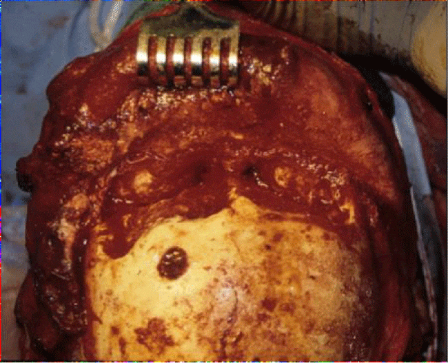
Figure 3:
Operative view of a patient undergoing the Riedel procedure.
-
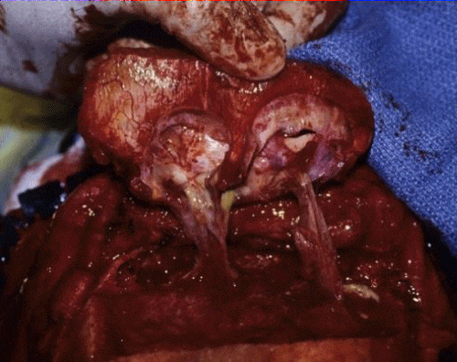
Figure 4:
Operative view of a patient undergoing the Riedel procedure showing multiple mucoceles eroding the anterior table of the frontal sinus.
• The flap is elevated to the supraorbital rims exposing the frontal bone and the area of osteomyelitis of the anterior table, which is often fragmented with a moth-eaten, chalky appearance, is removed with rongeurs. The bone is cut back to areas of active bleeding bone. This may entail removal of bone beyond the confines of the frontal sinus as involvement of the diploe permits extensive calvarial spread. Even after this, there may be microscopic foci of infection present that may eventually cause a recrudescence of the process. Similarly, abnormal areas of the posterior table of the frontal sinus should be removed.
• Following removal of the diseased bone, the sinus cavity is prepared as for an osteoplastic flap with all mucosa removed. The remaining bone of the posterior table is burred down to remove microscopic remnants of mucosa, which may follow the course of the veins of Breschet and result in secondary mucocele formation or intracranial spread. All bony ridges and septa are removed.
• The nasofrontal ducts are stripped of their mucosa and obliterated with temporalis fascia.
• The presence of supraorbital or other extramural ethmoid cells are exenterated, as are all other diseased sinuses.
• Any dural dehiscences or lacerations are repaired by primary suturing or fascial grafting.
• Cultures are taken from the operative site and the wound is copiously irrigated.
• The supraorbital ridges are exposed and reduced sufficiently to permit collapse of the forehead soft tissues into the defect and to contact the posterior table and any exposed dura to produce complete obliteration. The supraorbital and supratrochlear nerves are preserved if at all possible. Implicit in this is some degree of deformity, the severity of which depends of the prominence of the supraorbital ridges.
• A Penrose drain is inserted and the wound is closed in layers.
• With a significant degree of deformity of the forehead, a cranioplasty can be performed, generally with an alloplastic material (acrylic) or autogenous bone grafts, after an adequate disease free interval both clinically and on radionuclide evaluation.
Treatment
Acute frontal osteomyelitis requires drainage of the subperiosteal collection along with endoscopic or external drainage (trephine) of the frontal sinus. This is always performed in conjunction with prolonged IV antibiotic therapy. Imaging of the brain and orbits is essential because of the high incidence of intracranial and orbital complications associated with Pott’s puffy tumor which may also require neurosurgical and ophthalmologic intervention.
In the great majority of cases of acute and early chronic osteomyelitis, sinus drainage and intravenous antibiotics is curative. However, in a small subset of patients who are immunocompetent, with no or few comorbidities, the disease progresses to a refractory state. Failure to maintain nasofrontal outflow patency, persistent intracavity disease, failure to totally remove infected bone and secondary mucocele formation all contribute to the process.
The clinical history of patients with chronic osteomyelitis is of repeated courses of oral and IV antibiotics and an escalating level of aggressive surgical procedures. Multiple endoscopic frontal sinusotomies are general the initial treatment in these patients. This includes the DRAF III procedure which involves creating a single, large outflow tract for the bilateral frontal sinuses. In patients that fail endoscopic approaches, external procedures (i.e. frontal exploration and debridement, external frontoethmoidectomy, osteoplastic flap) are then employed. In a small subset of patients, all of these approaches fail to achieve a long term disease free state. When external procedures are performed the outcome is often compromised by insufficient removal of diseased bone to prevent cosmetic deformity. Antibiotic therapy also masks the smoldering infection which spreads through the bone.
In the pre-antibiotic era there are few papers addressing the management of chronic frontal osteomyelitis. Mohr and Nelson, in 1982, presented 4 cases (all male, mean age 33 years) secondary to trauma (2), chronic sinusitis (1) and mucocele (1). Clinical findings included: Pott’s Puffy tumor (2), orbital findings (2), fistula (1) and intracranial abscess (4) [12]. Surgical management was craniotomy and craniectomy in all 4 cases, cranialization in 2 cases, 1 in combination with the Riedel procedure and 2 cranioplasties. This small series demonstrates all the hazards in managing chronic frontal osteomyelitis, even with prolonged intravenous broad spectrum antibiotics. One patient required 3 craniotomies; the 2 cranioplasties failed; 1 patient developed symptoms 9 years after trauma; and 1 patient had bone involvement extending to the fronto-parietal suture line.
In 2002, Marshall and Jones reported 7 cases (6 male, mean age 39 years) of chronic frontal osteomyelitis secondary to frontal sinusitis [13]. All patients had a Pott’s puffy tumor, 2 sinocutaneous fistula, 3 a subdural abscess and 1 a periorbital abscess. All underwent bone debridement, 3 with craniotomy and 2 had a Riedel procedure. Complications included a recurrent brain abscess requiring craniotomy and sepsis with a subphrenic abscess and pericarditis.
Similarly, in the contemporary literature, there is very little mention of the Riedel procedure. Raghavan and Jones in 2004 reported 5 patients (3 male, mean age 55 years) treated with the Riedel procedure [14]. The clinical findings were Pott’s puffy tumor (3), fistula (4), periorbital collections (2) and epidural abscess (1). Collectively, these patients underwent numerous endoscopic procedures, 7 external frontoethmoidectomies, 5 frontal explorations and 4 osteoplastic flaps. The authors modified the procedure by chamfering the supraorbital ridges to reduce the cosmetic deformity, similar to Killian. No complications were reported after a follow up of 9 months to 6.5 years (mean 3.5 years).
External drainage is generally required as endoscopic ventilation has often proved to be inadequate. Concurrent intravenous antibiotic therapy is maintained for 4-8 weeks. The organisms cultured are diverse and include gram positive (Staphylococcus aureus, Streptococcus milleri), gram negative (Pseudomonas, E. coli, Enterococcus) and anaerobes (Peptostreptococcus, Bacteroides, Fusobacterium) [2,11,13,15]. The cultures are often polymicrobial and require broad spectrum antibiotic coverage, with the final selection based on intraoperative cultures.
Reconstruction of the operative defect is generally delayed for at least 1 year during which time the patient remains asymptomatic and radionuclide imaging (triphasic bone scan, indium white blood cell [WBC] scan) is negative. However, despite these precautions, the literature contains examples of cranioplasty failures. The 2 traumatic cases of Mohr and Nelson having a methylmethacrylate acrylic cranioplasty failed; in both cases the implant was placed after 1 year and became infected 2.5 and 4 years later and required removal [12]. Collet et al., repaired 3 cases of Pott’s puffy tumor developing 1-30 years following frontal reconstruction [15]. The authors recommend that primary reconstruction of the operative defect should not be performed at the time of the ablative procedure, especially with traumatic defects. In our series of 22 cases, both deferred cranioplasties failed.
Conclusion
1. Chronic frontal osteomyelitis is an indolent process that develops over many years in immunocompetent patients.
2. The natural history is generally of multiple progressively more invasive surgical procedures and numerous courses of antibiotic therapy.
3. Common presenting signs are the presence of Pott’s puffy tumor, periorbital cellulitis and cutaneous fistulae.
4. While acute frontal osteomyelitis may resolve with sinus ventilation and prolonged antibiotic therapy, chronic osteomyelitis requires more radical drainage and debridement of the diseased bone. Continued infections may require the Riedel procedure.
5. Despite its radical nature, the Riedel procedure may not be curative because of intraosseous spread beyond the frontal sinus and development of secondary mucopyoceles.
6. Patients with frontal osteomyelitis and Pott’s puffy tumor often have associated intracranial abscess from septic retrograde thrombophlebitis and may require craniotomy and craniectomy.
7. The bacteriology of frontal osteomyelitis is diverse and polymicrobial and requires prolonged wide spectrum systemic antibiotic therapy.
8. Cranioplasty should be delayed for at least 1 year with negative radionuclide imaging. Frontal reconstruction at the time of repair of traumatic frontal injuries carries a high failure rate.
-
-
- Pott P (1760) Observations on the nature and consequences of wounds and contusions of the head: fractures of the skull, concussions of the brain, &c. London: Printed for C. Hitch and L Hawes. 38-58. Link: https://goo.gl/FVVgNf
- Hicks CW, Weber JG, Reid JR, Moodley M (2011) Identifying and managing intracranial complications of sinusitis in children: a retrospective series. Pediatr Infect Dis J 30: 222-226. Link: https://goo.gl/xX2fhX
- Ibarra S, Aguirrebengoa K, Pomposo I, Bereciartua E, Montejo M, et al. (1999) Osteomyelitis of the frontal bone (Pott's puffy tumor). A report of 5 patients. Enferm Infecc Microbiol Clin 17: 489-492. Link: https://goo.gl/vgstBg
- Pender ES (1990) Pott's puffy tumor: a complication of frontal sinusitis. Pediatr Emerg Care 6: 280-284. Link: https://goo.gl/HtN4tp
- Tsai BY, Lin KL, Lin TY, Chiu CH, Lee WJ, et al. (2010) Pott's puffy tumor in children. Childs Nerv Syst 26: 53-60. Link: https://goo.gl/v6ZzpH
- Ketenci I, Unlu Y, Tucer B, Vural A (2011) The Pott's puffy tumor: a dangerous sign for intracranial complications. Eur Arch Otorhinolaryngol 268: 1755-1763. Link: https://goo.gl/xeE7Ey
- Nisa L, Landis BN, Giger R (2012) Orbital involvement in Pott's puffy tumor: a systematic review of published cases. Am J Rhinol Allergy 26: e63-70. Link: https://goo.gl/1Py3J6
- Blumfield E, Misra M (2011) Pott's puffy tumor, intracranial, and orbital complications as the initial presentation of sinusitis in healthy adolescents, a case series. Emerg Radiol 18: 203-210. Link: https://goo.gl/F2UBqq
- Bambakidis NC, Cohen AR (2001) Intracranial complications of frontal sinusitis in children: Pott's puffy tumor revisited. Pediatr Neurosurg 35: 82-89. Link: https://goo.gl/P9y69g
- Younis RT, Lazar RH, Anand VK (2002) Intracranial complications of sinusitis: a 15-year review of 39 cases. Ear Nose Throat J 81: 636-638, 640-632, 644. Link: https://goo.gl/sqexRi
- Akiyama K, Karaki M, Mori N (2012) Evaluation of adult Pott's puffy tumor: our five cases and 27 literature cases. Laryngoscope 122: 2382-2388. Link: https://goo.gl/2GoQMd
- Mohr RM, Nelson LR (1982) Frontal sinus ablation for frontal osteomyelitis. Laryngoscope 92: 1006-1015. Link: https://goo.gl/EnTzJS
- Marshall AH, Jones NS (2000) Osteomyelitis of the frontal bone secondary to frontal sinusitis. J Laryngol Otol 114: 944-946. Link: https://goo.gl/KV15aL
- Raghavan U, Jones NS (2004) The place of Riedel's procedure in contemporary sinus surgery. J Laryngol Otol 118: 700-705. Link: https://goo.gl/MMF5p6
- Verbon A, Husni RN, Gordon SM, Lavertu P, Keys TF (1996) Pott's puffy tumor due to Haemophilus influenzae: case report and review. Clin Infect Dis 23: 1305-1307. Link: https://goo.gl/iaxYqL
-


Table 1:
Patients Undergoing Riedel Collapse Procedure.*
Craniotomy