Authors:
Francesco Messina1, Francesco Loria2*, Giuseppe Loria3, Luciano Frosina4, Francesca Frosina5 and Enrico Morano2
1Department of Radiology PO Locri, ASP 5 Reggio Calabria, Italy
2Department of Radiology PO Palmi, ASP 5 Reggio Calabria, Italy
3Department of Radiology PO Lamezia Terme, ASP 5 Reggio Calabria, Italy
4School of Medicine, University of Messina, Italy
5School of Medicine, Campus Bio-Medico Roma, Italy
Received: 04 July, 2015; Accepted: 30 July, 2015; Published: 03 August, 2015
Francesco Loria, Department of Radiology PO Palmi, ASP 5 Reggio Calabria, Italy, E-mail:
Messina F, Loria F, Loria G, Frosina L, Frosina F, et al. (2015) Sonography and Contrast Enhancement MDCT in the Evaluation of Complicated Autosomal Dominant Polycystic Kidney Disease. Arch Renal Dis Manag 1(1): 003-007. DOI: 10.17352/2455-5495.000002
© 2015 Messina F, et al. This is an open-access article distributed under the terms of the Creative Commons Attribution License, which permits unrestricted use, distribution, and reproduction in any medium, provided the original author and source are credited.
Sonography; MDCT; ADPKD; MPR; Kidneys
Objectives: Our study is finalized to assess the role of Sonography and MDCT in the diagnostic work-up of patients with complicated Autosomal Dominant Polycystic Kidney Disease (ADPKD).
Methods: Thirty-five patients with ADPKD underwent Sonography, un-enhanced and contrast-enhancement MDCT for flank pain, haematuria and fever. Sonographic evaluation was made with patients in the supine position, full bladder, by making the patient with deep inalations, and other bedsores were used (side, prone) using other types of acoustic windows. MDCT studies were performed with volumetric acquisition technique (un-enhanced and contrast enhancement), and the relative images were evaluated at the appropriate work-station using MPR, cMIP, MIP thin and thick, and Volume Rendering (VRT) reconstructions. Two different Radiologists, with experience in genitourinary imaging, analysed image quality.
Results: All patients of our study had complicated cystic formations. The diameter of all cysts was valuated by Sonography and MDCT. Correlation between cystic diameters and Sonography/MDCT measurements was assessed using Pearson correlation test: tests were considered significant at P < 0.05. Cyst haemorrhage was present in all thirty-five patients, seen as high-density cysts, which were mostly bilateral. Sonography showed that these cysts had: sharply outlined contours (n = 7), sharp interfaces with adjacent renal parenchyma (n = 13), imperceptible walls (n = 9), and homogeneous density (n = 6), and all them did not enhance following e.v. contrast administration. The MDCT study also revealed, in addition to Sonographic results, the presence of: cyst infection (n = 13) cases, air within the infected cyst (n = 4), thickening and enhancement of peri- and paranephric fasciae (n = 5), abscesses in the posterior paranephric space and adjoining psoas muscle (n = 3), and cyst wall calcifications (n = 9). In 1 case MDCT revealed a soft tissue density enhancing mass in one of the cysts; this proved to be a renal cell carcinoma by the biopsy. We also identified renal calculi in 5 patients.
Results: All patients of our study had complicated cystic formations. The diameter of all cysts was valuated by Sonography and MDCT. Correlation between cystic diameters and Sonography/MDCT measurements was assessed using Pearson correlation test: tests were considered significant at P < 0.05. Cyst haemorrhage was present in all thirty-five patients, seen as high-density cysts, which were mostly bilateral. Sonography showed that these cysts had: sharply outlined contours (n = 7), sharp interfaces with adjacent renal parenchyma (n = 13), imperceptible walls (n = 9), and homogeneous density (n = 6), and all them did not enhance following e.v. contrast administration. The MDCT study also revealed, in addition to Sonographic results, the presence of: cyst infection (n = 13) cases, air within the infected cyst (n = 4), thickening and enhancement of peri- and paranephric fasciae (n = 5), abscesses in the posterior paranephric space and adjoining psoas muscle (n = 3), and cyst wall calcifications (n = 9). In 1 case MDCT revealed a soft tissue density enhancing mass in one of the cysts; this proved to be a renal cell carcinoma by the biopsy. We also identified renal calculi in 5 patients.
Introduction
ADPKD is a hereditary form of adult cystic renal disease, characterized by the unrelenting enlargement of innumerable cysts derived from renal tubules. ADPKD is one of the most common serious hereditary disease, found in 1:400 to 1:1000 individuals, and by far the most common hereditary cause of end stage renal failure (ESRF) [1]. The kidneys are normal at birth, and with time develop multiple cysts. Clinical presentation is variable and includes: dull flank pain of variable severity and time course (most common); abdominal or flank masses; haematuria; hypertension (usually develops at the same time as renal failure); renal functional impairment to renal failure. Macroscopically the kidney demonstrates a large number of cysts of variable size (from a few millimeters to many centimeters), filled with fluid of variable colours (from clear or straw colored to altered blood or chocolate coloured to purulent when infected) [2]. Besides monitoring renal function by standard measurements, follow-up of patients with ADPKD is based largely on radiologic investigations that are performed with Sonography, MDCT and Magnetic Resonance (MR), with the aim of evaluating renal cyst morphology and volume and estimating the amount of residual renal parenchyma [3]. The aim of this our study is to assess the role of Sonography and MDCT in the diagnostic work-up of patients with complicated ADPKD.
Materials and Methods
Patients selection
Thirty-five patients (Men = 25, Women = 10), with median age of 48 years old, with ADPKD, had been retrospectively subjected for a period of time of one year (April 2014 – April 2015) with Sonography, un-enhanced and contrast-enhancement MDCT for flank pain, haematuria, or fever.
MDCT examination
All MDCT examinations included were performed on a 32-slices multidetector row CT system (Light Speed, G.E.), using a low-dose protocol for kidneys and abdominal cavity, with volumetric acquisition technique, before and after the intravenous administration of the contrast, and the relative images were evaluated at the appropriate work-stations using MPR (axial, sagittal and coronal); cMIP; MIP thin and thick, and VRT reconstructions. The images were reconstructed as 1.2-mm-slices. MDCT examinations comprised the steps:
First step: unheanced-CT;
Second step: administration of contrast material at the rate 120ml 3-4 ml/sec, preceded by injection of furosemide/20mg and followed by infusion rapid drop in 250ml of saline solution. We made three phases:
Arterial phase (or Cortico-Medullary) performed at 30-35 seconds by the administration bolus of contrast agent;
Parenchimographic step: performed at 90-110 seconds the bolus of contrast agent;
Caliceal-Pielic phase (or excretory): performed at approximately 5-8 minutes infusion of contrast material;
Third step (or final step): Processing (Post-Processing) at dedicated MDCT work-station (Carestream Workspace). The Post-Processing parameters of cysts that were analysed in the study were: side, location, dimensions, and various complications (haemorrhage, infection, air within infected cyst, thickening and enhancement, abscess, density enhancing mass).
Sonography examination
Sonographic evaluation of all patients was carefully performed using a GE Logique Expert 5 equipment. The retroperitoneal position of kidneys imposed for an optimal exploration using a convex probe with a frequency of 3,5 MHz to obtain a good compromise between the capacity of penetration of the ultrasounds and the possibility of an adeguate resolution to get a complete anatomic detail. Patients were studied in the supine position, full bladder, by making the patient with deep inhalations. Other bedsores were used (side, prone) using other types of acoustic windows. The dorsal acoustic window uses for the exploration the axial and sagittal scans. Another window used is the anterior acoustic window. It had been widely used, because it appears very smooth and had always been used in the exploration of the right kidney (thanks to the acoustic window represented by the liver). The same parameters of Post-Processing MDCT were analysed in the Sonographic study.
Images analysis
Two different Radiologists, with experience in genito-urinary imaging, analysed image quality and blindly evaluated type, dimension and location of cystic lesions in these patients, the structure of kidneys and proximal urinary tracts and reported the diagnostic utility level for both examinations (Tables 1, 2). In particular, Radiologists analysed in detail the upper, middle and lower calyces, pelvis and proximal ureter on each side, and then they evaluated the images assigning independently a score in the diagnosis of renal cysts, according to a three-grades scale: score 1 indicated negative results (absence of disease); scores 2 indicated positive results (presence of disease); score 3 indicated that the presence or absence of cysts could not be determined.
-
Table 2:
Extra-renal sites of the cysts and their dimensions.
Statistical analysis
We had utilized the statistical software. Correlation between cystic diameters and Sonography/MDCT measurements was assessed using Pearson correlation test: tests were considered significant at P < 0.05.
Total diameters of renal cysts increased significantly during the MDCT valuation (P < 0.01) as a result of a significant increase/modification in cyst architecture (P < 0.01).
Results
All patients of our study had complicated cystic formations (Table 3). In 28 patients normal architecture of both renal parenchyma was greatly subverted by the presence of multiple, numerous and voluminous cysts, the largest of which presented maximum diameter of 15 centimeters (in the middle renal calyces), that “imprisoned” the kidneys, occupying almost the entire abdominal cavity (Figures 1, 4). The cysts had also diameter between 0,9cms and 4cms (in the upper renal calyces); between 1,3cms and 9cms (in the lower renal calyces); between 1cm and 5cms (in the renal pelvis), and between 0,6cms and 2cms at proximal urinary tracts. Other five patients showed cysts in the liver (Figure 2), with diameter between 0,5cms and 3cms. Two patients, respectively, had even cysts at the level pancreatic (dimensions between 0,3 and 1cm), and ovarian (diameter between 0,8cms and 2,5cms) (Table 4). In two patients were also highlighted, which exhibits accessories, aneurysms of the abdominal aorta (Figure 3). We divided all observed cysts at Sonography and contrast enhancement MDCT in five categories, depending on their presentation: 1) smooth walls, 2) fluid/corpusculated content, 3) internal septa, 4) calcifications or calcified rim; 5) tokens solids. Cyst haemorrhage was present in all thirty-five patients, seen as high-density cysts, which were mostly bilateral. Sonography showed that these cysts had (Table 5): sharply outlined contours (n = 7), sharp interfaces with adjacent renal parenchyma (n = 13), imperceptible walls (n = 9), and homogeneous density (n = 6), and all them did not enhance following e.v. contrast administration. The MDCT study also revealed (Table 6), in addition to Sonographic results, the presence of: cyst infection (n = 13) cases. Other findings included: air within the infected cyst (n = 4), thickening and enhancement of peri- and paranephric fasciae (n = 5), abscesses in the posterior paranephric space and adjoining psoas muscle (n = 3), and cyst wall calcifications (n = 9). In 1 case MDCT revealed a soft tissue density enhancing mass in one of the cysts; this proved to be a renal cell carcinoma by the biopsy. We also identified renal calculi in 5 patients. Our statistical analysis demonstrates that total diameters of renal cysts increased significantly during the MDCT valuation (P < 0.01) as a result of a significant increase/modification in cyst architecture (P < 0.01). The top-score resulted for MDCT was better than that of Sonography. In particular, the two dedicated-Radiologists found a significant relationship between that they observed at MDCT and complicated ADPKD.
-

Table 3:
Complications of cysts.
-
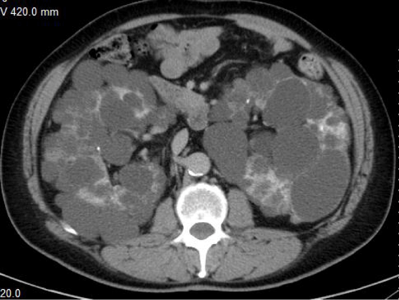
Figure 1:
Computed Tomography revealed that the normal architecture of both kidneys was subverted by the presence of innumerable, enlarged, voluminous cysts. Renal cysts were so huge that occupied almost the entire abdominal cavity, and the kidneys appeared to be “imprisoned” by them.
-
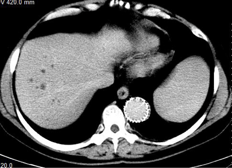
Figure 2:
Computed Tomography also showed many hepatic cysts, scattered throughout the various hepatic segments.
-
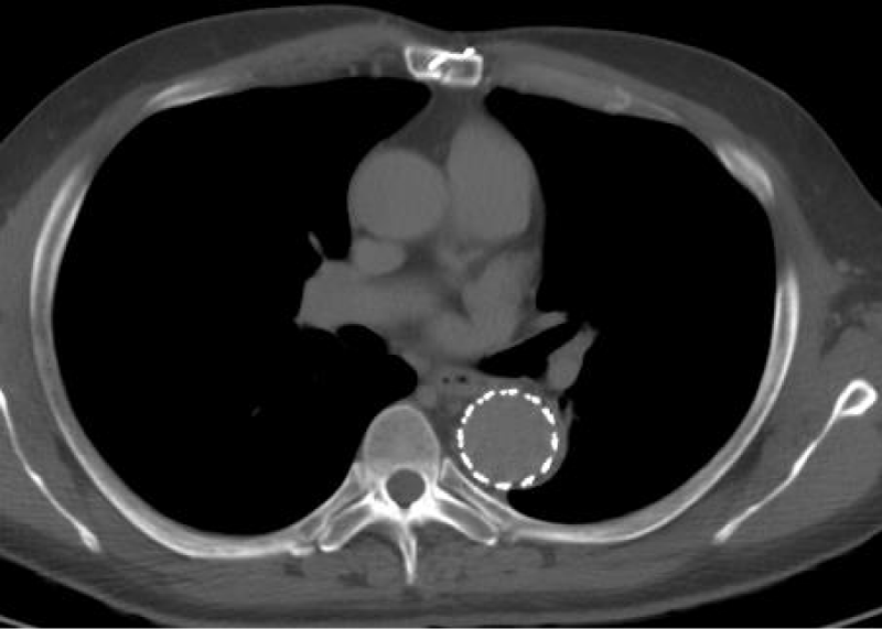
Figure 3:
There were no signs of endoleak of the thoracic endoprothesis.
-
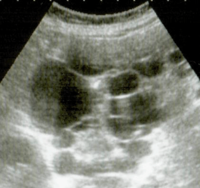
Figure 4:
Abdominal US showed the voluminous renal cysts.
-

Table 4:
Extra-kidneys sites of the cysts and their diameters.
-

Table 5:
Sonographic cystic results.
-

Table 6:
MDCT cystic results.
Discussion
ADPKD is caused by at least two (and possibly three) genes located on separate chromosomes. ADPKD-1 is due to a 14 kb transcript in a duplicated region on the short arm of chromosome 16, very near the a-globine gene cluster and the gene for one form of tuberous sclerosis. ADPKD-2 has been assigned to the long arm of chromosome 4 [4,5]. Cysts originate in renal tubules. Proliferation of tubule epithelial cells modulated by endocrine, paracrine, and autocrine factors is a major element in the pathogenesis of renal cystic diseases [6]. ADPKD is characterized by the unrelenting enlargement of innumerable bilateral cysts derived from renal tubules. This cystic growth often leads to a grotesque renal enlargement [7,8]. It is interesting that the largest cyst that we found had a diameter of 15 centimeters that imprisoned the kidneys, occupying almost the entire abdominal cavity. In our study we used Sonography and MDCT, and state of the art of Imaging Literature, to evaluate complicated ADPKD. In fact, relatively early in life, the cysts trigger secondary complications including renal infections, bleeding, stones, rupture of the cysts, pain, hypertension, hematuria and obstruction of the urinary tract. In particular, the formation of stones tends to occur in subjects with multiple and larger cysts. Renal insufficiency is usually not detected until the fifth or sixth decade of life; in the patients that developed renal insufficiency is very important monitoring the rate of GFR that is the most sensitive parameter in the evaluation of the disease progression. The rate of GFR decline is usually constant. The most important symptoms of ADPKD are: abdominal and lombar pain, hematuria, headache, dyspepsia, nicturia. ADPKD is a systemic disorder: cysts appear with decreasing frequency in the kidneys, liver, pancreas, brain, spleen, ovaries, and testis. Cardiac valvular disorders, abdominal and inguinal hernias, and aneurysms of cerebral and coronary arteries and aorta are also associated with ADPKD. Some reports have indicated that patients with ADPKD have aortic fragility; hypertension is also a known risk factor for aortic dissection [9,10]. Sonography, MDCT and MR have been used for many years to quantify the increase in renal volume in patients with ADPKD. Imaging with these techniques had also been used to accurately quantify the rate of increased kidney and total cyst volume in patients [11]. In our study we demonstrate that Sonography is a good choice for repeated imaging as it is fast, relatively inexpensive and lacks onizing radiation. It is able both to suggest the diagnosis and to assess for cyst complications. Simple renal cysts will appear anechoic with well-defined imperceptible walls, posterior acoustic enhancement (amplification) and lateral shadowing (extinction). Cysts with haemorrhage or infection will demonstrate echogenic material within the cyst, without internal blood flow. Calcification may develop. Renal cell carcinomas in contrast, although usually cystic in the setting of ADPKD, will have solid components of thick septa with blood flow. We think that Sonography of kidneys is the first imaging diagnostic method to detect ADPKD. The typical Sonography figure is generally bilateral, characterized by kidney size with increased echogenicity, with subverted structure by the presence of multiple cysts and spread to the whole body, of varying sizes, with the renal parenchyma poorly represented. Sonography is higly sensitive and specific in patients > 30 years of age, and it’s a non-invasive method, of low cost, easily repeatable and achievable [12]. MDCT is even more accurate than US identifying the ADPKD. It allows an accurate assessment of number, size, location and extension of renal cysts, and also detected in other organs (such as liver, spleen, and pancreas). MDCT can also well identified compression of adiacent abdominal structures, such as aneurysms and aortic dissections [13-16].
Conclusions
This our study on thirty-five patients with complicated ADPKD shows the importance of the preliminary assessment of these patients with Sonography, that provides valuable, preliminary diagnostic information, suggesting the initial diagnosis. The most complete characterization of complicated ADPKD is provided by MDCT: a combination of unenhanced and contrast-enhanced MDCT allows correct diagnosis and differentiation among the various complications affecting patients with ADPKD. MDCT can produce high quality images of the kidneys and the cysts. The entire abdomen cavity can be visualized during the same examination, and an accurate assessment of the number, size and extent of renal cysts, and also in other organs, is usually well demonstrated. In general, MDCT is an effective diagnostic method and also allows for more precise indications for subsequent treatment choices, playing a key-role among the various options.
-
-
-
- Gabow PA (1993) Autosomal Dominant Polycystic Kidney Disease. N Engl J Med 329: 332-342.
- Grantham JJ, Chapman AB, Torres VE (2006) Volume progression in autosomal dominant polycystic kidney disease: The major factor determining clinical outcomes: Clin J Am Soc Nephrol 1:148–157.
- Antiga L, Piccinelli M, Fasolini G, Ene-Iordache B, Ondei P, et al. (2006) Computed Tomography Evaluation of Autosomal Dominant Polycystic Kidney Disease Progression: A Progress Report. Clin J Am Soc Nephrol 1: 754-760.
- Wołyniec W1, Jankowska MM, Król E, Czarniak P, Rutkowski B (2008) Current Diagnostic evaluation of autosomal dominant polycistic kidney disease. Pol Arch Med Wewn 118: 767-773.
- Sessa A1, Ghiggeri GM, Turco AE (1997) Autosomal dominant polycystic kidney disease: clinical and genetic aspects. J Nephrol 10: 295-310.
- Martinez JR, Grantham J (1995) Polycystic kidney disease: etiology, pathogenesis, and treatment. Department of Medicine University of Medical Center, Kansas City 41: 693-765.
- Grantham JJ (1996) The etiology, pathogenesis and treatment autosomal dominant polycystic kidney disease: recent advances. Am J Kidney Dis 28: 788-803.
- Gagnadoux MF. Cystic kidney disease. Rev Prat (1997); 47: 1536-1540.
- Osawa Y1, Omori S, Nagai M, Obayashi H, Maruyama H, et al. (2000) Thoracic aortic dissection in a patient with autosomal dominant polycystic kidney disease treated with maintenance hemodialysis. J Nephrol 13: 193-195.
- Minami T, Sakamoto A (2009) Thoracic aortic dissection complicating autosomal dominant polycystic kidney disease: report of a case. Kyobu Geka 62: 924-927.
- Bae KT, Grantham JJ (2010) Imaging for the prognosis of autosomal dominant polycystic kidney disease. Nat Rev Nephrol 6: 96-106.
- Mostbeck GH1, Zontsich T, Turetschek K (2001) Ultrasound of the kidney: obstruction and medical diseases. Eur Rad 11: 1878-1889.
- Akira Kawashima, Terri J Vrtiska, Andrew J LeRoy, Robert P Hartman, Cynthia H. McCollough, et al. (2004) CT Urography. Radiographics 24: S35-S58.
- Van Der Molen AJ, Cowan NC, Mueller-Lisse UG, Nolte-Ernsting CC, Takahashi S, et al. (2008) CT Urography: definition, indications and techniques. A guideline for clinical practice. Eur Rad 18: 4-17.
- Tsushima Y (1999) Functional CT of the kidney. Eur Rad 30: 191-197.
- Chow et al. (2001) MDCT urography with abdominal compression and 3-reconstruction. AJR 177: 849-855.20:620-6.
-


Table 1:
Distribution of renal regions and dimensions of the cysts.